


 52 / 88

HIPERTENSIÓN / 2016 / VOL. 21
52
Los resultados de la intervención dependen de la causa de la hipertensión renovascular. La displasia
fibromuscular es más susceptible de éxito que la estenosis de arteria renal por ateroesclerosis. El éxito técnico
de la angioplastía oscila entre un 40 a un 90% y el éxito clínico entre un 50 a un 85%. La colocación de stents
tiene un éxito técnico del 95% y un éxito clínico primario de un 70 a un 80%
19
.
BIBLIOGRAFÍA
1.
Dworkin LD, Cooper CJ. Clinical practice. Renal-artery stenosis. N Engl J Med 2009; 361:1972.
2.
Textor SC, Lerman L. Renovascular hypertension and ischemic nephropathy. Am J Hypertens 2010; 23:1159.
3.
Zhender C. Hipertensión arterial renovascular. Rev. Med. Clin. Condes 2009; 20(3) 348 – 353.
4.
Gilles Soulez, Vincent L. Oliva, Sophie Turpin, et al. Imaging of Renovascular Hypertension: Respective Values of Renal Scintigraphy,
Renal Doppler US, and MR Angiography. RadioGraphics 2000 20:5, 1355-1368.
5.
Hirsch AT, Haskal ZJ, Hertzer NR, et al. ACC/AHA 2005 guidelines for the management of patients with peripheral arterial disease
(lower extremity, renal, mesenteric, and abdominal aortic): executive summary a collaborative report from the American Association
for Vascular Surgery/Society for Vascular Surgery, Society for Cardiovascular Angiography and Interventions, Society for Vascular
Medicine and Biology, Society of Interventional Radiology, and the ACC/AHA Task Force on Practice Guidelines (Writing Committee
to Develop Guidelines for the Management of Patients With Peripheral Arterial Disease). J Am Coll Cardiol. 2006;47(6):1239-1312.
6.
Williams GJ, Macaskill P, Chan SF, et al. Comparative accuracy of renal duplex sonographic parameters in the diagnosis of renal
artery stenosis: paired and unpaired analysis. AJR Am J Roentgenol 2007; 188:798.
7.
Radermacher J, Chavan A, Bleck J, et al. Use of Doppler ultrasonography to predict the outcome of therapy for renal-artery stenosis.
N Engl J Med 2001; 344:410.
8.
Zachrisson K, Herlitz H, Lönn L, et al. Duplex ultrasound for identifying renal artery stenosis: direct criteria re-evaluated. Acta
radiológica 2016.
9.
Echevarría JJ, Miguélez JL, López-Romero S, et al. Arteriographic correlation in 30 patients with renal vascular disease diagnosed
with multislice CT. Radiologia 2008; 50:393.
10. Glockner JF, Vrtiska TJ. Renal MR and CT angiography: current concepts. Abdom Imaging 2007; 32:407.
11. ESUR Guidelines in contrast media, Versión 8.1. Disponible en:
http://www.esur.org/guidelines/sp/index.php12. FDA Drug Safety Communication: New warnings for using gadolinium-based contrast agents in patients with kidney dysfunction.
Disponible en:
http://www.fda.gov/Drugs/DrugSafety/ucm223966.htm13. Wang Y, Alkasab TK, Narin O, et al. Incidence of nephrogenic systemic fibrosis after adoption of restrictive gadolinium-based
contrast agent guidelines. Radiology 2011; 260:105.
14. Pedersen EB. New tools in diagnosing renal artery stenosis. Kidney Int 2000; 57:2657-77.
15. Thadhani RI, Camargo CA, Jr, Xavier RJ, Fang LS, Bazari H. Atheroembolic renal failure after invasive procedures: natural history
based on 52 histologically proven cases. Medicine (Baltimore). 1995;74(6):350-358
16. Lao D, Parasher PS, Cho KC, Yeghiazarians Y. Atherosclerotic Renal Artery Stenosis—Diagnosis and Treatment. Mayo Clinic
Proceedings. 2011;86(7):649-657. doi:10.4065/mcp.2011.0181.
17. Derkx FH, Schalekamp MA. Renal artery stenosis and hypertension. Lancet 1994; 344:237.
18. Martin L.G., Rundback J.H., Wallace M.J. et al. Quality Improvement Guidelines for Angiography, Angioplasty, and Stent Placement
for the Diagnosis and Treatment of Renal Artery Stenosis in Adults. (2010) Journal of Vascular and Interventional Radiology, 21
(4) , pp. 421-430.
19. Iqbal S, Sharma A, Wicky ST. Arterial interventions for renovascular hypertension. Semin Intervent Radiol. 2009 Sep;26(3):245-52.
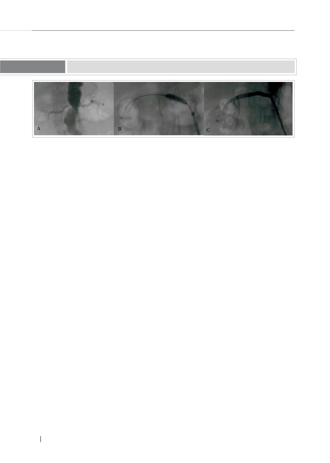
Estenosis de arteria renal derecha (A) tratada con stent. (B) Instalación de stent. (C) Control postinstalación
Figura Nº 7